
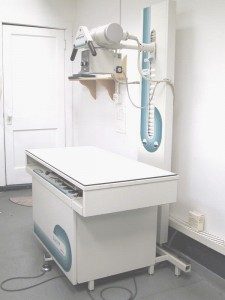
 There are 3 components to the radiology area of our clinic; the x-ray machine, the automatic x-ray processor, and the viewer. X-rays are generally performed by animal health technologists (AHT) and veterinary assistants. Animal health technologists have received extensive training in how to properly take x-rays. We will walk you through the process of taking an x-ray:
- The animal is safely restrained and positioned on the x-ray table by an animal health technologist and veterinary assistant. Usually the animal is awake or given mild sedation to take an x-ray. For certain x-rays (ex. hips and skull), the animal needs to be given a general anesthetic in order to obtain proper positioning. The AHT and assistant wear protective lead coverings while taking radiographs.
- The x-ray film is taken into the darkroom, labeled appropriately and put into the automatic x-ray processor for development. The radiographs only take about one minute in the processor.
- The animal health technologist initially views the x-ray on a viewer (a machine that projects light) to determine if the technique and positioning was acceptable. If it is, the veterinarian interprets the radiograph and makes a diagnosis.
- The x-ray is sent to a veterinary radiologist via courier for a second opinion.
Call us to book an appointment (403) 262-3237